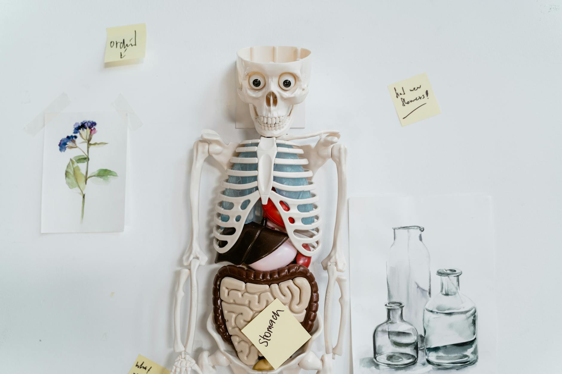
Studying human anatomy can feel like a huge challenge. With hundreds of terms for bones, muscles, and organs, it’s easy to get overwhelmed. But what if learning could be more engaging, creative, and even relaxing? Enter anatomy coloring pages, a powerful and fun tool for students of all ages.
Coloring isn’t just for kids. See the therapeutic benefits of coloring for adults. When you color in a diagram of the heart or the brain, you’re engaging multiple senses. This process helps build strong visual and kinesthetic connections, making it much easier to remember complex information compared to just reading text. Let’s explore how you can use this simple technique to master anatomy.
Getting Started with Anatomy Coloring
Before we tour the human body, let’s cover the basics of using anatomy coloring pages as a study aid. Understanding how to use them effectively can transform your learning experience from a chore into a creative journey.
What Exactly Is an Anatomy Coloring Page?
An anatomy coloring page is a black and white illustration of an anatomical structure, complete with labels, designed specifically for learning. Unlike a simple drawing, these pages are organized to teach you about the body’s systems. The act of coloring helps reinforce the location, shape, and function of each part as you spend time focusing on it. Whether you’re a high school biology student or a future doctor, these pages offer a practical way to learn. And with modern tools, you can even create custom diagrams on any topic you need.
How to Use Anatomy Coloring Pages for Maximum Learning
To get the most out of your coloring session, a little strategy goes a long way. Make sure you’ve got the right pencils and fineliners with this guide to choosing the right coloring tools for your project. The best method is to assign a unique color to each anatomical structure and use that same color for its corresponding label. For example, if you color the eye’s lens red, you should also color the word “lens” in red. This simple trick creates a powerful mental link between the term and its visual representation. For more practice ideas, explore our coloring tips and techniques. This technique helps you build a mental map of the body, making it significantly easier to recall information later.
Finding Free Printable Anatomy Coloring Pages
You don’t need to buy expensive books to get started. A free printable anatomy coloring page is a fantastic resource you can download and print at no cost. Many educational websites offer collections of high quality PDF or PNG files covering essential diagrams like the heart, brain, and skeleton. These are perfect for students and teachers who need study materials in a pinch. Try this quick tip for creating unique coloring activities for your classroom. Plus, web‑based generators like CustomColoringPages.com host huge libraries of free pages and even let you create new ones instantly (here’s how AI crafts custom coloring pages in seconds), so you can always find the exact diagram you need.
What Is an Anatomy Coloring Book PDF?
An anatomy coloring book PDF is a digital book that combines many coloring sheets into a single, convenient file. These are often organized by body system and include detailed illustrations with labels. While many popular books are available for purchase, you can often find free PDF collections online. Kenhub, for example, offers a free PDF with six detailed pages covering tricky structures like the brain, skull, and heart. A PDF format is great because you can print pages as many times as you like or even color them digitally on a tablet.
Exploring the Body’s Systems: A Tour Through Coloring
Now, let’s grab our coloring pencils and take a journey through the major systems of the human body.
The Circulatory System in Color
The circulatory system is the body’s transportation network. Coloring its components helps you trace the path of blood and understand how every cell gets the oxygen it needs.
- Heart Coloring Page: The heart is central to our anatomy, and a heart coloring page is the perfect way to learn its four chambers, major vessels, and valves. As you color the atria, ventricles, aorta, and vena cava, you’ll start to visualize how blood flows through this powerful pump. Did you know a healthy adult heart pumps about 7,500 liters of blood every day?
- Pulmonary Circulation Coloring Page: This specific diagram shows the loop of blood flow between the heart and lungs. You can use one color for the pulmonary arteries carrying oxygen poor blood to the lungs and another for the pulmonary veins carrying oxygen rich blood back to the heart. This helps clarify that pulmonary arteries are the only arteries carrying deoxygenated blood.
- Blood Vessel Coloring Page: A general blood vessel coloring page can teach you the difference between arteries, veins, and capillaries. Arteries have thick, muscular walls, while veins are thinner and have valves. A fun fact: if you laid out all the blood vessels in an adult, they would stretch over 60,000 miles.
- Abdominal Aorta Branch Coloring Page: The abdominal aorta is the main artery in your abdomen, and it has several major branches. Coloring the celiac trunk, mesenteric arteries, and renal arteries helps you remember which organs each branch supplies with blood.
- Artery of Head and Neck Coloring Page: This page focuses on the carotid and vertebral arteries that supply blood to the brain. As you color, you can trace the path of the internal carotid artery to the brain and the external carotid artery to the face.
- Artery of Upper Limb Coloring Page: See how blood travels from your shoulder to your fingertips. You’ll color the subclavian artery as it becomes the axillary artery and then the brachial artery, which is the same vessel used for taking blood pressure.
- Artery of Lower Limb Coloring Page: Follow the blood supply to the legs, starting from the external iliac artery which becomes the femoral artery in the thigh. Coloring this pathway helps you understand how major muscles and bones in the leg get their oxygen.
Coloring the Digestive System
From the first bite to the final exit, the digestive system is a long and winding road. Coloring it piece by piece makes it much easier to learn.
- Mouth, Pharynx, and Esophagus Coloring Page: This diagram covers the start of the journey. You can color the tongue, teeth, and the epiglottis, a flap that cleverly prevents food from going down your windpipe.
- Digestive Tract Coloring Page: Get the big picture with a diagram of the entire digestive tract. A fascinating fact is that the gastrointestinal tract is about 25 to 30 feet long in an adult. You can color the stomach, the 20 foot long small intestine, and the 5 foot long large intestine.
- Large Intestine Coloring Page: This page zooms in on the colon. You can color the cecum, appendix, and the different sections (ascending, transverse, descending). The large intestine is home to over 700 species of bacteria that help us produce certain vitamins.
- Liver Coloring Page: The liver is the body’s largest internal organ and a metabolic powerhouse, performing over 500 vital functions. Coloring its lobes, the gallbladder tucked underneath, and its unique dual blood supply helps you appreciate its complexity.
- Tooth Anatomy Coloring Page: A cross section of a tooth reveals its layers: the super hard enamel, the softer dentin, and the living pulp inside. Enamel is the hardest substance in the human body. As you color, you’ll see why a cavity can become painful if it reaches the inner layers.
Mapping the Skeletal System
The skeleton provides our body’s framework, and coloring is a fantastic way to learn the 206 bones that make it up.
- Skeleton Coloring Page: A full skeleton coloring page is the perfect starting point. A cool fact is that we are born with about 270 bones, but many fuse together as we grow, leaving adults with 206. More than half of your bones are in your hands and feet.
- Skull Coloring Page: The skull is a complex puzzle of 22 bones. A skull coloring page helps you learn the names and locations of the frontal, parietal, occipital, and facial bones. Coloring the “strangely shaped bones” of the skull makes learning them much faster.
- Vertebral Column Coloring Page: Learn the different regions of the spine: cervical, thoracic, and lumbar. A popular mnemonic to remember the number of vertebrae is to think of meal times: breakfast at 7 (cervical), lunch at 12 (thoracic), and dinner at 5 (lumbar).
- Scapula Bony Feature Coloring Page: The scapula, or shoulder blade, has many bumps and grooves where muscles attach. Coloring features like the acromion (the bony tip of your shoulder) and the glenoid cavity (the socket for your arm bone) helps you understand how the shoulder moves.
- Bone of Upper Limb Coloring Page: This page focuses on the 30 bones in each arm, from the humerus in the upper arm to the radius and ulna in the forearm and the many small bones of the wrist and hand.
- Bone of Lower Limb Coloring Page: Similarly, each lower limb contains 30 bones. You can color the femur, which is the longest and strongest bone in the body, as well as the tibia, fibula, and the bones of the foot.
Understanding Joints Through Coloring
Joints are where bones meet, and coloring them clarifies how they move.
- Joint Coloring Page: Learn the difference between immovable fibrous joints (like skull sutures) and freely movable synovial joints (like the knee). A diagram of a synovial joint will show the articular cartilage, joint capsule, and synovial fluid that allow for smooth movement.
- Knee Joint Coloring Page: The knee is one of the most complex joints. A detailed coloring page will show the ligaments that provide stability, like the anterior cruciate ligament (ACL) and posterior cruciate ligament (PCL), as well as the cushioning menisci.
Visualizing the Muscular System
Coloring muscles helps connect their anatomy to their actions.
- Muscle of Face Coloring Page: The face has over 20 flat muscles that create our expressions. You can color the zygomaticus major, the “smiling muscle,” and the powerful masseter muscle used for chewing.
- Muscle of Neck, Chest and Thorax Coloring Page: Learn about the sternocleidomastoid in the neck that turns your head, the pectoralis major on your chest, and the intercostal muscles between your ribs that help you breathe.
- Muscle of Arm and Forearm Coloring Page: Differentiate the biceps brachii on the front of the arm from the triceps brachii on the back. You’ll also see the groups of muscles in the forearm that control your wrist and fingers.
- Muscle of Thigh and Hip Coloring Page: Color the four muscles of the quadriceps group on the front of the thigh and the three hamstring muscles on the back. You’ll also find the gluteus maximus, one of the body’s strongest muscles.
- Muscle of Leg and Foot Coloring Page: This page highlights the calf muscles (gastrocnemius and soleus) that connect to the strong Achilles tendon. You’ll also see the muscles on the front of the leg that lift your foot.
The Nervous and Endocrine Systems
These two systems control everything in the body. Coloring helps demystify their complex structures.
- Nervous System Structure and Function Coloring Page: Get an overview by coloring the Central Nervous System (brain and spinal cord) one color and the Peripheral Nervous System (nerves) another. This visually separates the control center from the communication network.
- Neuron Coloring Page: A neuronal coloring page shows the parts of a nerve cell, including the dendrites, cell body, and axon. You can color the myelin sheath that insulates the axon and allows nerve impulses to travel incredibly fast, up to 268 miles per hour.
- Brain Coloring Page: A brain coloring page simplifies one of the body’s most complex organs. The human brain contains around 86 billion neurons. Coloring the different lobes (frontal, parietal, temporal, occipital) helps you remember their locations and primary functions.
- Brain Structure and Brain Part Coloring Page: These pages zoom in on specific areas. You might color the cerebellum, which coordinates balance, or internal parts like the corpus callosum, which connects the brain’s two hemispheres.
- Cranial Nerve Coloring Page: There are twelve cranial nerves that emerge directly from the brain. Coloring each one and its pathway is a great way to memorize their names and functions, from the olfactory nerve for smell to the facial nerve for expressions.
- Endocrine Gland Coloring Page: This page shows the hormone producing glands, like the pituitary “master gland” in the brain, the thyroid in the neck, and the adrenal glands on the kidneys. Coloring them helps you map out this vital chemical messaging system.
Coloring the Respiratory System
Follow the path of air from the nose to the lungs.
- Respiratory System Coloring Page: This diagram shows the entire pathway, including the nasal cavity, larynx (voice box), trachea (windpipe), and bronchi. Coloring the diaphragm muscle below the lungs helps you understand how breathing works.
- Lung Coloring Page: A detailed lung coloring page shows the lobes of the lungs (three on the right, two on the left). It also illustrates the tiny air sacs called alveoli, where gas exchange happens. Humans have about 300 million alveoli, providing a massive surface area for oxygen to enter the blood.
The Sensory Organs in Detail
Our senses connect us to the world. Coloring these intricate organs helps you understand how they work.
- Eye Coloring Page: An eye coloring page reveals the parts that allow us to see. You can color the cornea, iris, lens, and the light sensitive retina at the back of the eye.
- Ear Coloring Page: An ear coloring page separates the outer, middle, and inner ear. You’ll color the eardrum, the three tiny ossicle bones (the smallest in the body), and the cochlea, the spiral shaped organ for hearing.
- Sense Organ of Skin Coloring Page: The skin is our largest sensory organ. A cross section shows the different receptors for touch, pressure, pain, and temperature embedded in its layers.
- Olfactory Organ Coloring Page: Learn about the sense of smell by coloring the olfactory epithelium in the nasal cavity. This is where millions of receptor neurons detect odors and send signals to the brain.
- Tongue Coloring Page: The tongue is covered in papillae that contain our taste buds. A diagram will show the different types of papillae and the nerves responsible for taste.
- Tooth Anatomy Coloring Page: A detailed look at a tooth helps you understand its structure. As you color the layers of enamel, dentin, and pulp, you build a strong visual memory of tooth anatomy.
Create Your Own Custom Anatomy Pages
Sometimes, you need a very specific diagram that’s not in a standard coloring book. Maybe it’s a particular view of the knee joint or a close up of a neuron’s synapse. Instead of spending hours searching, you can create exactly what you need in seconds. With a tool like CustomColoringPages.com, you can simply type a description to generate a unique coloring page or upload a photo to convert it into line art (here’s how to transform your photos into fun coloring pages in seconds).
Ready to create your own unique diagrams? Generate your own anatomy coloring pages in seconds and build a personal library perfectly tailored to your studies.
Frequently Asked Questions about Anatomy Coloring Pages
Are anatomy coloring pages effective for medical students?
Yes, absolutely. Many medical students use anatomy coloring pages as a supplementary study tool. For a broader overview, see Adult Coloring Books: Benefits, Tools & Beginner Tips. The act of coloring engages active learning, which can improve memory retention of complex structures, especially for highly detailed areas like the skull or cranial nerves.
Can I make my own anatomy coloring pages?
Of course. If you can’t find the exact diagram you need, you can use an online generator. Websites like CustomColoringPages.com allow you to create a coloring page from any text description, giving you full control over your study materials.
What’s the best way to color these pages for learning?
For the best results, use a consistent color‑coding system. Assign a specific color to each structure and use that same color for its label. When you want to add depth and contrast, try these color blending techniques. This creates a strong visual association between the term and its location.
Where can I find specific diagrams, like a neuron coloring page?
You can find them in specialized anatomy coloring books, on educational websites, or by generating them yourself. A quick search for “free printable neuron coloring page” will often yield good results from educational sources.
Do I need a special book or can I find free pages online?
You don’t need to buy a book to start. There are thousands of free, high quality anatomy coloring pages available online for immediate printing. These are perfect for targeted study sessions on specific topics.